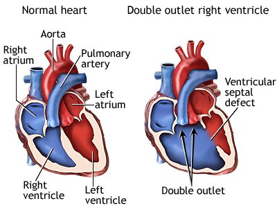
What is double outlet right ventricle?
In double outlet right ventricle (DORV), a unique condition which is characterized by attachment of both great arteries ( aorta and pulmonary ) to right ventricle. Left ventricle ejects blood through ventricular septal defect. ( Check Normal heart for more information).
Due to this arrangement , there is compulsory mixing of pure and impure blood. DORV is associated with many other defects which decide clinical manifestation. Associated defects include atrial septal defect, atroventricular canal defect, tricuspid atresia, pulmonary stenosis, aortic stenosis, coarctation of aorta etc. Baby can present like 1) large VSD with excessive blood flow to lung ; 2) pulmonary stenosis: TOF like cyanosis, blue baby 3) TGA: transposition of great arteries, 4) coarctation etc.
Signs and symptoms of double outlet right ventricle
Depending upon associated condition, DORV can present in ways mention above. The symptoms can present themselves even in early neonatal period.
- Cyanosis: Blue or purple tint to lips, skin and nails
- Poor eating and poor weight gain
- Rapid breathing or shortness of breath
- Profuse sweating,
- More sleepiness than normal
- Heart murmur
Testing and diagnosis of DORV
- Fetal Echo: Heart defect can be diagnosed before birth when baby is in mother’s womb. It helps to plan delivery at well equipped centre. Parents can also make informed decision.
- Electrocardiogram(EKG or ECG):
- Echocardiogram: ( refer to Paediatric 2D echocardiography)
- Chest x-ray
- Cardiac MRI and Ct scan:
- Cardiac catheterization
Treatments for double outlet right ventricle
A few babies may require medication after birth to stabilize after birth. Surgery is only definitive option available. Depending upon nature of anatomy ( structure of heart) variety of surgeries are planned. In a few cases, with help of tunnel ( baffle) , left ventricle is connected to aorta . It prevents mixing of oxygenated and de-oxygenated blood. Associated anomalies like coarctation fo aorta, ASD, PDA, pulmonary stenosis are also addressed at the same time.
Those with anatomy like TGA, require arterial switch operation. In this case, VSD is closed and position of aorta and pulmonary artery is interchanged.
In a few cases, it not possible to create baffle, close VSD or one of the ventricle is very small, a special type of approach called as single ventricle palliation ( Fontan) is undertaken. It requires 2 to 3 stage surgeries.
Outlook and follow up care for DORV
Most babies after surgery live a healthy and good quality of life. A small subset may develop complication after over period of follow up. These are unique to underlying anatomy and nature of surgery like , baffle obstruction, pulmonary regurgitation, abnormal rhythm, coarctation, etc. They can be tackled with additional surgery or cardiac intervention.
Kids who underwent single ventricle palliation, forms a special group and need long term expert care. Kindly refer to Fontan for more discussion.
